
- Mourad – Biomedical Analysis
Dr. Mourad’s practical and hands-on teaching, research, development and commercialization activities inform his contributions to the presently proposed work. Dr. Mourad has extensive experience in mentoring undergraduate students in his interdisciplinary research, which involves applied mathematics and data analysis as well as clinical and lab-based research methodology. His undergraduate students have contributed as authors of over 33 published articles. The projects proposed here are similar in nature to the past projects that have so successfully involved undergraduates in research.
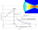
 Brain stiffness imaging. In [63] we expanded on a published method to improve brain imaging with ultrasound. Specifically, we used a commercial device that generates and tracks shear waves, each facilitated by ultrasound, to make maps of brain-tissue stiffness. In our published work we used a mouse model of stroke. In [62] we worked with two rodent models of focal traumatic brain injury. To make this technique work for human brain we need to work beyond the commercial device, to create our own algorithms for tracking the propagation of shear-waves through human-sized brain tissue. For this project, students will perform research that supports generation and gathering of data as well as its analysis, from research ultrasound devices applied to living animal brain. Specific mathematical analysis will include development of two-dimensional cross-correlation techniques to track ultrasound induced shear-wave propagation (whose speed is proportional to the shear modulus of tissue), development and testing of first principle models of that propagation as a function of material properties, and comparison of direct measures of brain-tissue stiffness (collected via use of calibrated indentation machines) with ultrasound-derived estimates of brain-tissue stiffness.
 Detecting vasospasm with ultrasound. After head trauma many patients bleed within or adjacent to their brain. This can cause the major blood vessels that feed to brain to contract (undergo spasm), with a result in reduced blood flow to the brain. Reduced blood flow reduces the delivery of oxygen to the brain, with eventual neuronal disability and death. We have a new means of collecting data on these blood vessels from patients within our Level One Trauma Center here at the University of Washingtons Harborview Medical Center, where Dr. Mourad has one of his academic appointments. For this project, students will analyze that data in its various forms using statistical methods, analysis of temporally involving images, feature extraction, etc, in order to improve the ability of this device to present to the physician in real time an image of the blood vessel in spasm, in real time. If successful, this project will lead to development of a device that will remove the need to move patients to brain-imaging devices, replacing their use with a device deployable at the patient bedside, without moving the patient.


You must be logged in to post a comment.